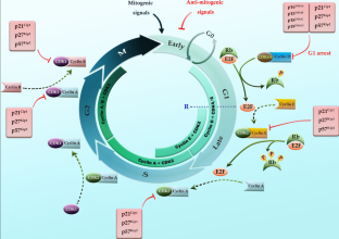
Journal of Cancer Research and Biology Diagrams Cell cycle regulation. At the end of mitotic metaphase: cyclin B level degradation begins resulting in lower amount of active MPF which brings about anaphase, telophase cytokinesis and eventually the cells reenters interphase.In summary, High levels of active MPF stimulate G2/M progression or mitosis whereas low levels favour return to interphase.DNA damage is the major reason that prevents
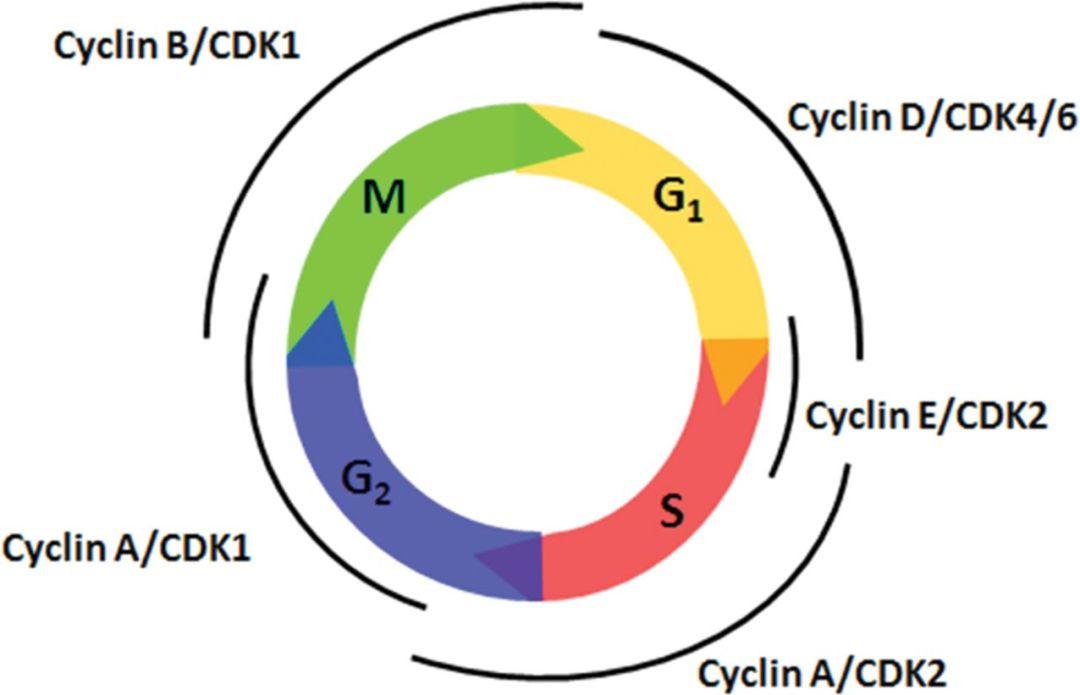
All cyclins are named according to the stage at which they assemble with CDKs. Common classes of cyclins include G 1 -phase cyclins, G 1 /S-phase cyclins, S-phase cyclins, and M-phase cyclins.

The cell cycle and its regulation by cyclins, CDKs, and CDKIs. The cell ... Biology Diagrams
Download scientific diagram | The cell cycle and its regulation by cyclins, CDKs, and CDKIs. The cell cycle is divided into four distinct phases (G 1 , S, G 2 , and M). The progression of a cell

The different cyclins and Cdks bind at specific points in the cell cycle and thus regulate different checkpoints. Figure \(\PageIndex{1}\): Activation of Cdks: Cyclin-dependent kinases (Cdks) are protein kinases that, when fully activated, can phosphorylate and activate other proteins that advance the cell cycle past a checkpoint. To become
Cell Cycle Regulation: Cyclins and CDKs Biology Diagrams
Cyclins Determine the Activity of CDKs (named because their levels change during the cell cycle). Cyclins are divided into four classes defined by their presence and activity during the cell cycle: G1 Cyclins (D cyclins) G1/S cyclin (Cyclin E) S-phase cyclins (cyclins E and A) M-phase cyclins (B cyclins) G1 cyclins (Cyclin D) coordinates the Cyclins and CDKs as in charge molecules of cell cycle progression, therefore their negative regulator actions are in focus in many cancer mechanism (García-Reyes et al., 2018). CDKs having role Download scientific diagram | 9: Overview of the CDKs and the cyclins. Each cyclin binds to one or several CDKs, which phosphorylate several different substrates. Cyclin-CDKs regulate
The cyclins, cyclin-dependent kinases (CDKs), CDK inhibitors (CKIs), and checkpoint proteins are examples of these internal signals that keep an eye on cellular parameters like cell growth, chromosome alignment, and DNA integrity. Protein kinases are the enzymes that activate or inactivate other proteins. They do these by phosphorylation.
